
Diagnostic equipment
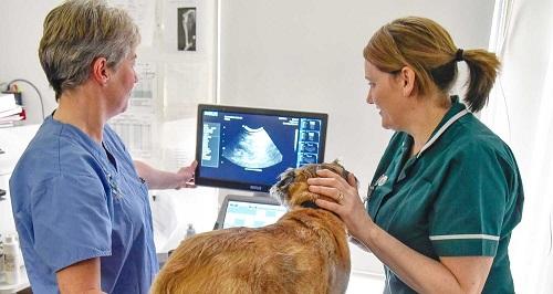
Ultrasonography is a diagnostic imaging technique which allows us to see an image of the inside of the body via reflected high frequency sound waves.
Ultrasound scans can be used to evaluate soft tissues, such as abdominal organs and the heart, but are not effective at imaging harder tissue like bone.
This technique is not invasive and involves applying a scanner probe to the body wall with the application of some ultrasound gel.
Sedation is often required to keep the pet still whilst they have their scan and some fur will need to be clipped to ensure good contact with the ultrasound scanner.
X-rays are a versatile diagnostic imaging technique. They are taken by passing a small amount of radiation through the animal onto an x-ray plate, which produces an image.
Having an x-ray taken is not painful, but sedation is often required as the animal needs to stay very still.
X-rays can provide a large amount of information for a wide range of conditions, including:
-
Orthopaedic disorders, for example broken bones and arthritis
-
Thoracic x-rays can help to assess the heart and lungs
-
Abdominal x-rays can be helpful when investigating cases of vomiting and diarrhoea, for example if bloated organs or a gastrointestinal blockage are suspected
-
Dental x-rays to assess whether a tooth needs removing
Endoscopy is another discipline which we are using more and more to aid clinical diagnosis. Our portfolio of scopes includes a large diameter flexible endoscope for gastro-intestinal purposes, a narrow diameter bronchoscope for respiratory system examination, and a rigid rhinoscope for nasal and urinary examination. Endoscopic examination of the body cavities is also achievable with our laparoscopic (keyhole surgery) equipment.